Neural Stem Cell Facility

About

The Neural Stem Cell Facility at CHINTA is at the forefront of human stem cell research, focusing on hiPSC-derived neurons and astrocytes and their implications for neurological disorders across human lifespan. At the centre of this endeavor lies the limitless capacity of stem cells to transform into various cell types, offering novel insights into human biology and its disease mechanisms.

Neural Stem Cell Facility

Resources

Our state-of-the-art lab adheres to international standards and provides a controlled environment for stem cell research. Equipped with clean rooms, biosafety hoods, tri-gas incubators, brightfield microscopes, and centrifuges, the facility supports reliable culturing and maintenance of neuronal and glial stem cells. These resources enable researchers to generate, expand, and study human-derived stem cell models with precision and reproducibility. This cutting-edge technology is used to screen novel compounds that can modulate neural stem cell behavior and function, aiming to develop groundbreaking treatments for neural disorders.

Latest finding

In vitro SCA12-patient derived brain organoids at CHINTA recapitulate neurodegeneration.

Behavioral Facility

About

Understanding animal behavior is fundamental to uncovering how the brain processes information, adapts to the environment, and generates complex functions. Our Behavioral facility serves as a hub for system-level neuroscience, enabling researchers to link cellular and circuit-level mechanisms with observable actions by integrating advanced tools with rigorous experimental design.

Resources

We house a comprehensive range of behavioral setups to study fear learning and memory, anxiety-related behavior, spatial navigation, reward-based learning, decision-making, social learning and pain-related behaviour. These include paradigms such as fear conditioning apparatus, elevated plus maze, open field, linear track setup, object location task, social interaction test, hot plate test and electronic Von Frey test.

Latest finding

Enhanced norepinephrine signaling in the ACC drives the affective-motivational and somatosensory responses to neuropathic pain.

Wet Biology Facility

About

The wet biology facility specializes in performing different types of molecular and biochemical analysis, which forms the backbone of neurological research. This facility executes several foundational assays for a wide range of research projects aimed at understanding the complexities of the nervous system and developing potential treatments for neurological conditions.

Resources

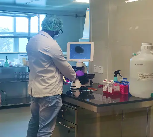
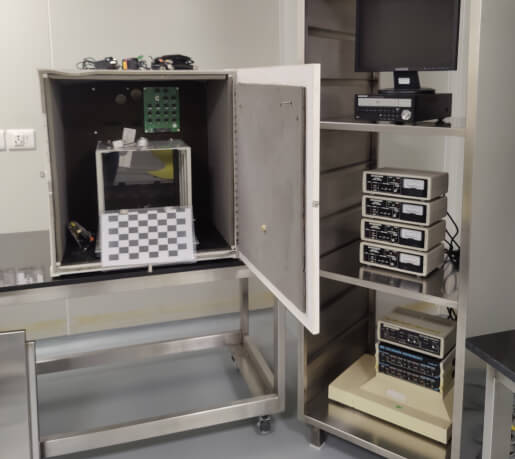
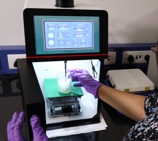
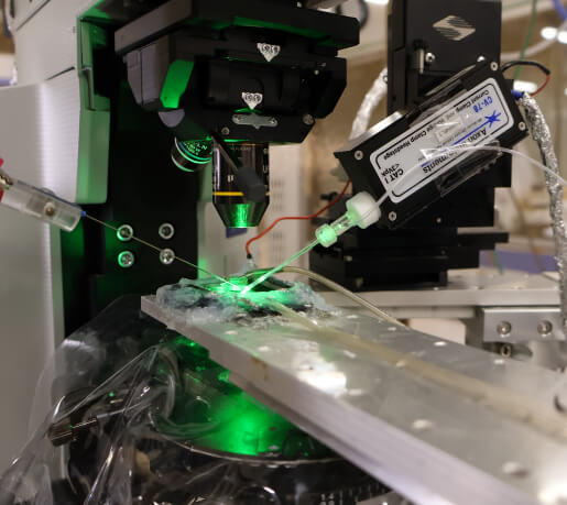
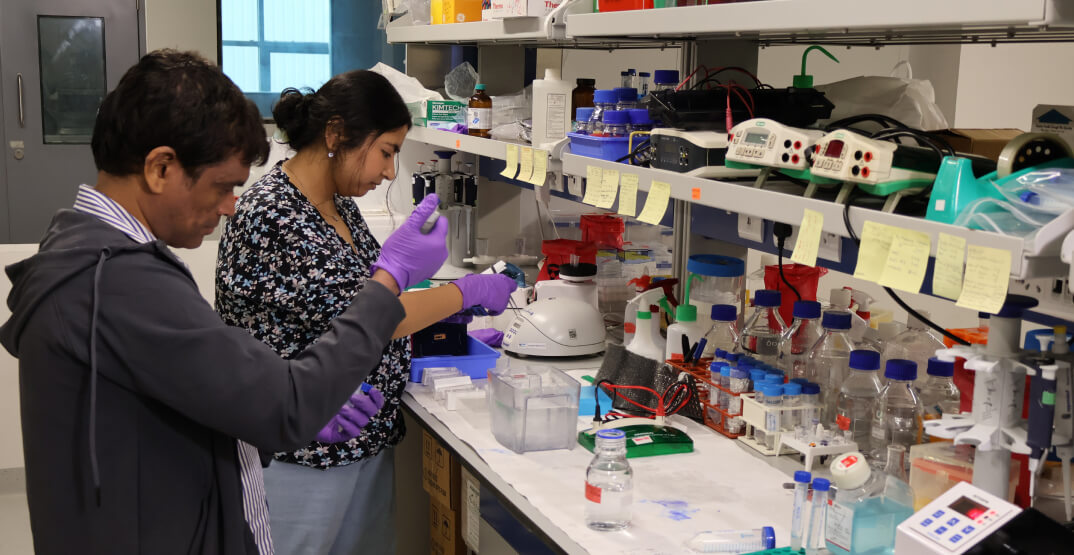
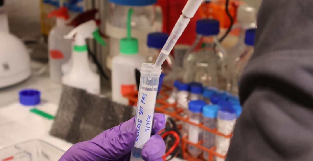
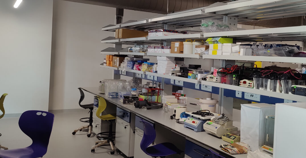
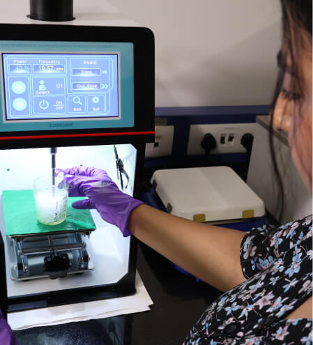
Leveraging state-of-the-art equipments, including Ultracentrifuge, RealTime PCR System, Multimodal microplate reader, Nanodrop spectrophotometer, Thermocyclers, Vibratome, Cryostat, Laminar flow hood, among others, the lab space is optimized for advanced molecular biology applications, such as tissue staining, immunophenotyping, quantitative RTPCRs, and other experiments involving DNA/RNA and cell-biological analyses.

Latest finding

Our efforts have generated novel Spino Cerebellar Ataxia clones to mimic the human disease in animal models

Neurophysiology Facility

About

Electrophysiological technologies allow researchers to study single-cell and network activity and can provide crucial insights into the functioning of individual neurons, circuits, and their roles in different behaviors. The experiments conducted in our neurophysiology facility bridge the gap between cellular and circuit-level neuroscience, significantly contributing to our understanding of brain function and disorders.

Resources

Our neurophysiology facility is equipped with multiple electrophysiology recording stations, enabling scientists to conduct single-cell, brain slice, and in vivo recordings. Additionally, the facility features vibratome machines for brain slice preparation and pipette pullers for creating glass electrodes with precise tips for patch-clamp experiments. The facility also includes an in vivo electrophysiology setup and stereotaxic implantation for recording electrical activity from behaving animals.

Latest finding

BTSP induction in hippocampal CA1 neuron occurs more robustly in males than in females, where the plasticity varies with estrous phases.

Imaging Facility

About

Visualizing neuronal structure and molecular dynamics is essential for understanding brain function at every level, from fundamental mechanisms to disease processes. At CHINTA, the imaging facility serves as a cornerstone for both basic and translational research, enabling scientists to capture dynamic cellular and molecular events with precision.

Resources

Our imaging facility has multiple advanced imaging setups. It has a dedicated laser scanning confocal microscope that allows high resolution functional imaging of live brain cells and fixed tissue. CHINTA has also built an advanced two-photon fluorescent lifetime microscopy setup for deep tissue imaging- the first of its kind in India. The imaging facility also includes epifluorescence and bright-field microscopes to understand different neuronal functions.

Latest finding

Successful activation of AMPK sensor in response to calcium influx visualized using two-photon time lapse imaging.